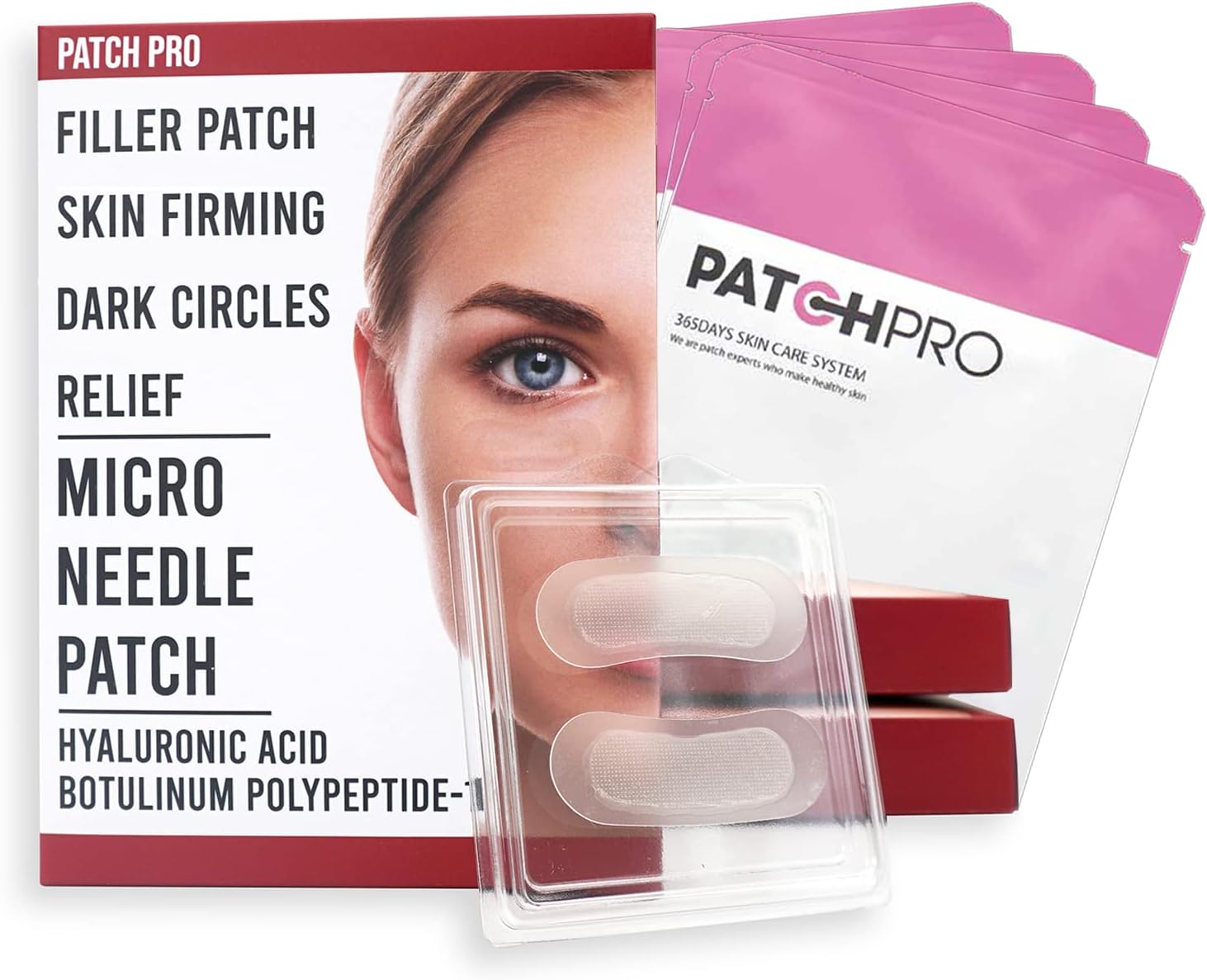
PATCH PRO MICRO NEEDLE PATCH 8pcs, self-dissolving microneedle patch, facial anti-wrinkle patches, crosslinked hyaluronic acids for forehead lines, smile line, fine line, eye wrinkle, Puffy eyes
FREE Shipping
PATCH PRO MICRO NEEDLE PATCH 8pcs, self-dissolving microneedle patch, facial anti-wrinkle patches, crosslinked hyaluronic acids for forehead lines, smile line, fine line, eye wrinkle, Puffy eyes
- Brand: Unbranded

Description
The main benefit of microneedle patches is obviously that they’re targeted to a specific area of skin, but they’re also safe. Since they cause no trauma to the skin, you won’t feel a thing (aside from a satisfying tingle indicating that they’re working) and they won’t cause any bleeding unlike dermarollers which are considerably more aggressive. Other manufacturing processes for microneedle fabrication include injection moulding [ 61], wet chemical etching [ 75], reactive ion etching [ 2, 76], hot embossing [ 4– 5], laser drilling [ 77], lithography plus electroforming [ 78– 79], drawing lithography [ 80– 81], two-photon polymerization [ 5, 82], and 3D printing [ 83– 84]. To date, DRIE of silicon; micromoulding; photolithography; and Lithographie, Galvanoformung, Abformung or lithography, electroplating, moulding (LIGA), using deep X-ray lithography, are the most extensively used manufacturing technologies for microneedle fabrication, although fabrication of longer microneedles (>400 μm) is difficult with some of these methods, notably DRIE. Drawing lithography has been used for high aspect ratio microneedles of heights 1600, 1200, and 600 μm [ 80– 81]. This technique involves spin coating of a viscoelastic thermosetting polymer such as SU-8 epoxy resin, followed by thermal curing and controlled drawing of the material in liquid form. It has so far been limited to low density arrays with relatively large spacing between adjacent microneedles (>900 μm). Animal studies suggest that the immune response to the flu vaccine may be stronger when delivered by a microneedle patch rather than by injection. However, larger clinical studies will be needed to assess the approach in people. Prausnitz has co-founded a company called Micron Biomedical that hopes to move the microneedle patch technology forward. “We now need to follow this study with a phase II clinical trial involving more people,” Prausnitz says, “and we hope that will happen soon.” Related Links Microneedling your skin can help active ingredients reach beyond the outer layer of skin, helping them to be more effective.
Delivery: Progress and Challenges - PMC Microneedles in Drug Delivery: Progress and Challenges - PMC
Actuating grafts appears to turn on cell signals related to the growth of new blood vessels and nerves; a promising finding for restoring mobility in muscle lost through disease or trauma.Hollow microneedles contain a lumen or internal channel for pressure-driven fluid communication through the microneedle and the skin [ 12, 54]. The fluid can be a drug, vaccine, blood, or ISF. This design enables the transporting of drug solutions and vaccines rather than their dehydrated form as is the case for dissolving microneedles or the competing needle-free methods, including powder jet delivery [ 55]. Solid microneedles are simpler to manufacture than hollow microneedles, so most preliminary studies were performed on solid versions. For solid microneedles, the vaccines or drugs are either coated on the microneedle surface or applied to the skin after micropores have been formed by the insertion of microneedles [ 12]. Therapeutics pre-coated on solid microneedles may dissolve off them after insertion into the skin. Gas-jet dry coating [ 56], liquid methods including repeated immersion and dip-in coating [ 57– 58], and spray coating [ 56, 59] are some of the techniques used for coating microneedle arrays with drugs and vaccines (mainly water soluble). Another approach for drug delivery using solid microneedles is to fabricate them entirely from biodegradable or water-soluble dissolving polymers. Drugs are encapsulated into the microneedle body and released as the microneedles dissolve [ 34, 37]. The main limitation of dissolvable microneedles is the limited choice of drugs that can be encapsulated into dissolvable polymer or polymer–sugar combinations [ 34, 36], and the short time and low volume of drug delivery. Dissolvable polymer microneedles of soluble poly(lactic- co-glycolic acid) (PLGA) and PLGA–polyvinylpyrrolidone (PLGA–PVP) layered combinations have been used to provide controlled drug delivery of bovine serum albumin (BSA), rather than instantaneous release [ 60]. The promise of vaccine MAP technology, and its likely impact on global health, is compelling; however, the challenge is now to bridge the gap between early clinical development and commercial reality. There are challenges in several areas including: Editor’s note: This article has been updated to clarify that this research was developed to help avoid preventable deaths in parts of the world where paper or digital systems for storing patients’ vaccination records aren’t available. Many vaccines require multiple doses spaced out at certain intervals; without accurate records, people may not receive all of the necessary doses. The method is still in an experimental stage and is not being used for any current vaccinations, including Covid-19 vaccines. However, with the advent of some spectacular science, microneedle patches that can be safely used at home are now available. They’ve been based on the same innovative skin health technology as microneedling using a dermaroller, but are even better and less invasive… Do Microneedle Patches Work for Acne?
An overview of microneedle applications, materials, and
Tate, J. E., Burton, A. H., Boschi-Pinto, C. & Parashar, U. D., World Health Organization-Coordinated Global Rotavirus Surveillance, N. Global, regional, and national estimates of rotavirus mortality in children <5 years of age, 2000-2013. Clin. Infect. Dis. 62, S96–S105 (2016). All immunogenicity results were analyzed by Prism software version 7 (GraphPad, CA, USA). Comparisons among individual samples were done using an unpaired t test. Comparisons among multiple groups were done using a two-way ANOVA. p< 0.05 was considered significant. Reporting summaryThe researchers showed that their new dye, which consists of nanocrystals called quantum dots, can remain for at least five years under the skin, where it emits near-infrared light that can be detected by a specially equipped smartphone. Antigen casting solutions for second-generation dMNP were prepared at lower vaccine concentrations while keeping the ratio of vaccine to excipient constant. To fabricate full dose IRV dMNP, casting solutions had a concentration of 0.625 mg/ml IRV, 2% w/v sucrose (VWR, OH, USA) and 0.4% w/v sodium methylcellulose. IPV casting solutions had a concentration of 5.8, 1.1, and 5.2 DU/μl of types 1, 2, and 3, respectively, 1.5% w/v maltodextrin (Sigma-Aldrich, MO, USA) and 0.5 % w/v xylitol. Full-dose combination patches were prepared to deliver 5 μg IRV and 40, 8, and 32 DU of IPV types 1, 2, and 3, respectively. Solutions for manufacturing the quarter dose dMNP contained lower vaccine doses while keeping the excipient concentrations constant. After deposition of the polymer matrix solution, all MN arrays were dried at 35 °C overnight. Stability studies Two or three doses of standalone IPV or combined IRV-IPV in the first-generation dMNP at full or half dose induced protective titers (≥1:8) of NA to the corresponding PV types 1, 2, and 3 in rats (Fig. 5d–f). The differences in antibody titer between post-vaccine doses 2 and 3 of full dose standalone IPV and full IRV-IPV were not statistically significant. Similarly, the differences in antibody titer between post-vaccine doses 2 and 3 of full-dose IPV, full-dose IRV-IPV, and half-dose IPV or half-dose IRV-IPV were not statistically significant. Of the three IPV types, IPV 2 was the most immunogenic and induced high geometric mean titers (GMTs) to PV type 2 even with a single dose. Some people described faint redness in the area of their skin where the patch had been as well as mild itching that lasted two to three days. But overall, the researchers found that giving the flu vaccine by the microneedle patch was safe, with no serious related side effects. More than 70% of those receiving the patch said they would prefer the flu vaccine patch over the flu shot or nasal spray flu vaccine. “This bandage-strip sized patch of painless and dissolvable needles can transform how we get vaccinated,” says NIBIB Director Dr. Roderic I. Pettigrew. “A particularly attractive feature is that this vaccination patch could be delivered in the mail and self-administered. In addition, this technology holds promise for delivering other vaccines in the future.”
Microneedle drug delivery - Wikipedia
CDC-9, a human G1P[8] RV strain, was cultivated in Vero cells and triple-layered particles (TLPs) and double-layer particles (DLPs) were purified from cell supernatants by using CsCl gradient centrifugation 38. The ratio of TLPs and DLPs was approximately 9:1 as measured by protein concentration with a Bradford assay. TLPs and DLPs in 50 mM HEPES, 150 mM NaCl, 5 mM CaCl 2 [pH 7.2-7.5] supplemented with 7% D-sorbitol were inactivated at 62 °C for 6 h and protein concentration was determined by Pierce™ Coomassie Bradford protein assay kit (Thermo Fisher Scientific, IL, USA) before being used for the fabrication of dMNP. Mono-bulks containing approximately 900, 450, and 650 DU/ml of IPV types 1, 2, and 3, were donated by GlaxoSmithKline, and were concentrated approximately 150, 100, and 75-fold by volume, respectively, and suspended in 0.375 mM histidine buffer (J.T. Baker ®, PA, USA). Mono-bulk concentration and buffer exchange were performed using Amicon Ultra centrifuge spin filters with 100 kDa MW cutoff at 4 °C, 4000 × g (Millipore Sigma, MA, USA). D-antigen contents were determined by ELISA 39. Dissolving microneedle patch (dMNP) fabrication Causing a deliberate injury to the skin to gain a benefit sounds a bit crazy, we’ll be the first to admit that. But when we look at the science behind microneedling, it all becomes clear. Nair, N. et al. VP4- and VP7-specific antibodies mediate heterotypic immunity to rotavirus in humans. Sci. Transl. Med. 9, eaam543 (2017). To improve manufacturing and delivery efficiency, second-generation dMNP, composed of 163 700-µm-tall MNs with a total MN volume of 2.7 µl, were fabricated similarly with the following differences. For the IRV-containing dMNP, the CMC in the formulation was replaced with methylcellulose (MC) and CMC was removed from the polymer matrix solution of IPV-containing dMNP. Since CMC is unable to be sterilized by filtration, methylcellulose (MC) was evaluated in combination with the other excipients and found to be a suitable replacement for CMC in the formulation to be more compatible with future GMP manufacturing. In addition, the combination IRV-IPV dMNP were fabricated as a single patch and not assembled halves of two separate arrays, as done with the first-generation combination dMNP. Dissolving microneedle patches were fabricated from polydimethylsiloxane (Dow Corning, MI, USA) molds using a two-step casting method (i.e., first an antigen-containing solution followed by a polymer matrix solution).Extensive research has recently been carried out on design, fabrication, and applications of microneedle systems. Microneedle patches, for example, could bring significant benefits from both patient and health professional perspectives due to reduced discomfort and enhanced convenience of application. In comparison to other microporation techniques such as ultrasound, thermal, electroporation, high-pressure needle-free injection, and lasers, microneedle-based systems are attracting favourable interest from both research and industry sectors. Microneedles have proved to be pain free, traversing the SC of the skin and penetrating the viable epidermis without stimulating nerve fibres. Microneedles may be integrated into biosensors, micropumps, microfluidic chips, and microelectronic devices.
- Fruugo ID: 258392218-563234582
- EAN: 764486781913
-
Sold by: Fruugo